
This page is grossly outdated and will be revamped soon. To learn about currently active research projects in the group see Publications.
We are lucky to live in exciting times.
Introduction of fluorescent proteins and the following rapid advance of microscopy methods precipitated a revolution in cell biology – suddenly we can "see" phenomena that few anticipated to find inside biological cells. These are emergent self-organized structures, such as spatial gradients, oscillations and propagating waves. What until recently was only a subject of theoretical hypotheses, now can be studied experimentally with the ever-expanding set of genetic, pharmacological and, most recently, chemical-genetic tools. Complexity of biological systems has long called for computational modeling. With the ever-increasing precision and specificity of experimental perturbations, we can now test and build our models iteratively, in close collaboration with experimentalists.
In our group we work with data generated by live-cell fluorescence imaging and a variety of biophysical techniques, such as FRAP, FRET and FCS, to build and parameterize mechanistic models of spatio-temporal structures emerging inside cells. We are lucky to stand on the shoulders of giants: in our work we can rely on the treasure-trove of theoretical concepts and methods developed over the decades by nonlinear physics and chemistry. There are more projects in our group than could fit on this page, so only a few can be covered in some detail. However the overarching questions we address in our research are the following:
How do biological cells break spatial symmetry? How do cells control location, size and number of nascent spatial structures? What are the common and evolutionary conserved biophysical mechanisms of intracellular morphogenesis?
Early morphogenesis of the baker’s yeast bud
Budding yeast, rather than dividing in the middle, proliferates by building daughter cells from scratch, on the side of the mother cell. The earliest manifestation of this is the emergence of the cluster of activated Rho GTPase Cdc42 (see below the movie courtecy of S. Okada and E. Bi, UPenn). We proposed a first cytoskeleton-independent biophysical mechanism for the self-organized emergence of the Cdc42 cluster that required neither upstream landmarks, nor actin-mediated vesicle transport. Our model, based on the idea of coupling of the membrane-cytoplasmic shuttling and autocatalytic activation of Cdc42 , predicted potential simultaneous emergence and the following competition between multiple Cdc42 clusters. This prediction was later confirmed together with Prof. D. Lew, Duke University .
Together with Prof. Erfei Bi, UPenn, we set out to investigate the emergence of the budding yeast septin ring, next event after the Cdc42 cluster formation in the cascade of amazing morphogenetic transitions leading to the formation of a new yeast cell. It was already shown that Cdc42 cluster is directly responsible for the emergence of the septin ring, yet, how Cdc42 recruits septins and why they form a ring, remained a puzzle.
In this project we unexpectedly discovered that septins recruited to the Cdc42 cluster inhibit the activity of Cdc42, most likely via Cdc42 GAPs. This negative feedback could be best seen under the conditions when polarized exocytosis, secretion of membrane vesicles within the cluster, was suppressed pharmacologically or genetically causing a novel “chasing” behavior of septins. Our model was able to explain this astounding jumping behavior via the competition of Cdc42 clusters for the effector-septin complexes (see the movie on the right).
Our modeling suggested that Cdc42-directed polarized exocytosis could hollow the septin density deposited in the middle of Cdc42 cluster via a physical insertion of membrane (see movie). This hypothesis received a strong support in the experiments of our collaborators.
Despite much effort by the research community, many questions related to the emergence of cell polarity and early bud morphogenesis remain unclear and controversial. Some of the questions we are interested in are:
What are the relative roles of actin-dependent and independent pathways in polarity establishment? How is the Cdc42 module regulated and what processes does it regulate? How is the polar distribution of proteins along the mother-daughter axis controlled at the later cell cycle stages?
Establishment and maintenance of polarity axis in fission yeast
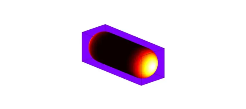
Fission yeast is another excellent model eukaryotic system to study cellular morphogenesis. Geometrically almost perfect, pill-shaped (more correctly, spherocylindrical) cells of this unicellular fungus can grow from both ends and strictly maintain one axis of polarity throughout essentially the entire cell cycle. However, under some genetic perturbations and cell stresses, fission yeast builds branches, which represent additional axes of polarized growth. Genetic analysis found that, in wild type cells, so-called polarity landmarks, set and maintained by the dynamic microtubules, keep memory of the location of zones of polarized growth.
Together with Prof. Ken Sawin, we performed a detailed kinetic analysis and proposed a model of the dynamics of this polarity landmark using live-cell imaging and extensive FRAP data. We demonstrated that the landmark is being continuously slowly rebuilt by the microtubules. Our modeling suggested a mechanism that both safeguards the polarity axis from the sudden short-lived perturbations and also auto-focuses the landmark preventing it from undesirable spatial spreading and loss of precision.
Many questions, however, remain open and call for detailed investigation. Thus, the role of Cdc42 GTPase has been only recently appreciated by the fission yeast community. Some of the questions we plan to address are:
How is the polarity axis created de-novo and maintained in fission yeast cells? What is the role of the Cdc42 cluster in fission yeast cell polarity? What are the common and divergent mechanisms of polarity regulation between the budding and fission yeast systems?
This article was published on